Cavities
Published on Aug 09 by Daniel Guidera under Cosmetic, Dentistry
What is a cavity? This is a common question in a dental office. In retrospect, this should have been the topic of my first post.
Earlier this week a gentleman came in with a cavity in his lower left wisdom tooth. The tooth already had a composite (white) filling on the biting surface, which was intact. I can’t take credit for it; this was the first time this patient had been in my office for restorative care. If you look closely on the X-ray, you can see a shadow on the left side of the tooth adjacent to the porcelain crown.
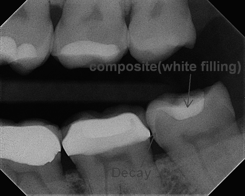
X-ray showing tooth decay
This raises the question “what is a cavity”? Now I am not a “bumper sticker” kind of guy, but if I was, I would have one that says “decay happens”. It’s just one of those things. However, in this case, I would venture to guess that when the crown on the adjacent tooth was done, the wisdom tooth was nicked by the drill, promoting decay. It isn’t necessarily carelessness by the dentist. You try to avoid nicking, but it happens. If it happens in my office I like to polish the enamel and place a fluoride varnish.
We could discuss the benefit of extracting vs. repairing the wisdom teeth, but that is for a later time. Also, the existing filling was composite. Removing a perfectly good filling only creates a bigger hole which makes the tooth weaker. Therefore, I chose to dovetail a new composite filling into the existing one.
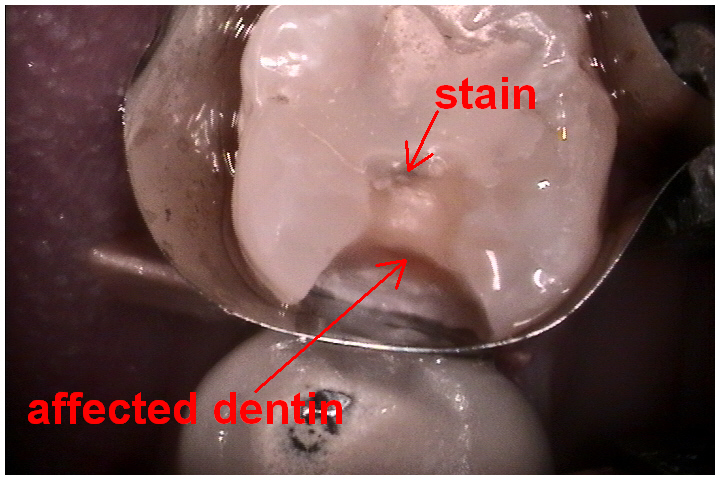
Stain & affected dentin
The first picture beautifully displays what decay is all about. Bacteria on the tooth eats simple carbohydrates and produces acid as a result. It’s the product of what is known in biochemistry as glycolysis.
The acid etches (decalcifies) the enamel, breaks down the enamel structure – allowing bacteria to reside deeper in the tooth while producing more acid. As the destruction pierces through the hard enamel, it breaks out and spreads into the softer dentin layer. From here it’s a race to the center of the tooth where the nerve resides.
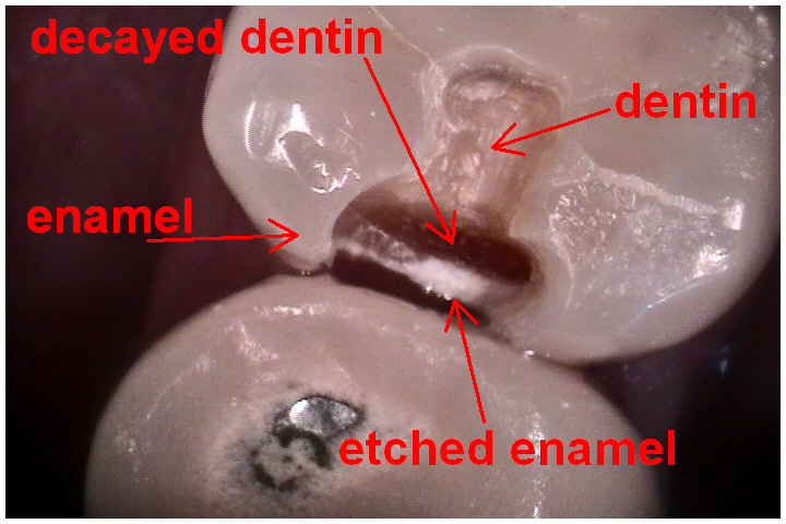
Drilled tooth
The next picture shows the decay removed and the tooth ready to be filled. Early on, color and texture are valuable guides in determining the presence of decay. As you get deeper into the tooth, color becomes less of an indicator. Notice some darker areas that are marked. Sometimes the tooth can be affected by the acid at the forefront of the decay and stained, but not infected and broken down.
These stains are very hard with clear borders. Why not remove the stain to play it safe? Don’t forget, bigger holes weaken teeth, and there is a nerve about 2 millimeters away that does not like being messed with!

Glass ionomer base
The next picture shows a base layer that I probably did not have to use in this case, but it has some chemical-physical characteristics that make it nice to use. It isn’t hard enough to use as a surface material in adult teeth.
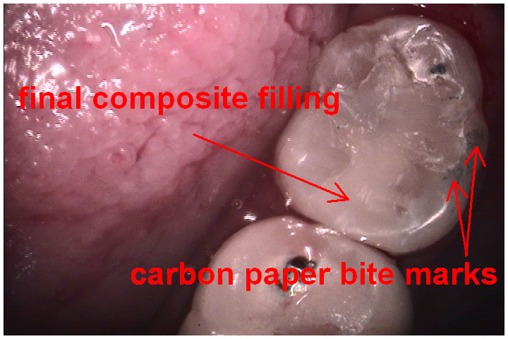
Final filling
The last picture shows the final composite filling. The part I anticipate, as a dentist, is the sound and feel of the dental floss at the end. If the floss glides through smoothly that means there’s an “open contact”. With this, food will get stuck between the teeth and cause problems.
But in this case the floss goes “SNAP!!!!” My work here is done.